How small...is small?
Steven Schultz
Princeton NJ -- Imagine a hair, a single strand of the thinnest, finest human hair. Slice it lengthwise into 10 strips. Each would be too small to see with the naked eye and would be about as wide as a typical cell, about 5 millionths of a meter.
|
|
|
|
|
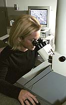
Senior Jennifer Shultz is
using the Imaging and Analysis Center for her
independent research comparing neurons in different
sized animal brains.
|
Objects this small could never show up in a conventional microscope. Ordinary light is too crude a medium to carry such delicate information. Light waves wash past atomic-scale features, like ocean waves rolling over seashells.
Electron microscopes, as the name suggests, work with electrons instead of light. Electrons move with a wavelength vastly smaller than that of visible light, so they can interact with correspondingly smaller objects.
An electron microscope focuses a beam of electrons onto the object, which spews out its own electrons and X-rays. The microscope creates an image by reading the energy and distribution of these emissions. One kind of microscope, the scanning electron microscope, sweeps the beam over the surface of an object. Another kind, called transmission electron microscope, creates a beam so strong it pierces thin materials and reveals structure within.
Interpreting these blasts of electrons requires not only sophistication in the machinery, but great experience and knowledge of physics by the operator.
|
|
|
|
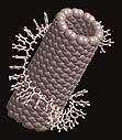
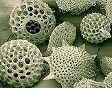
The carbon nanotube at left,
magnified about 25 million times, is made of a
single layer of carbon atoms and has attracted
attention for its strength and flexibility. The
image above depicts a single-celled algae magnified
about 1,000 times.
|
|
In this atomic realm, another major challenge is preparing the miniscule slide of material to be viewed. One device in the imaging lab cuts materials into slices just 40 millionths of a millimeter thick. Jennifer Shultz, an undergraduate studying brain cells -- a relatively enormous thing to view under an electron microscope -- estimated that preparing a brain sample takes her 10 days of solid work.
For Yao, all the difficulties are well worth the challenge.
"You have a wonderful tool here," he said. "You can shoot anything you want if you are interested." Quoting Ernst Ruska, the German engineer who invented the electron microscope in 1931, Yao said, "The light microscope opened the first gate to the microcosm. The electron microscope opened the second. What will open the third?"
See related article
Big picture begins with
smallest details
|
[an error occurred while processing this directive] |